Smart Blood Concentrates

The core focus on our business is based on Smart Blood Concentrates from Dr. Joseph Choukroun – the pioneer and inventor of PRF (Platelet Rich Fibrin) – who since 2001 has pushed the limits and research on the possibilities to advance post surgical wound healing and pain management.
Today his work is materialised as the A-PRF™ fibrin membrane solution and the i-PRF™ injectable PRF solution offering healthcare professionals a wide range of capabilities – all biologically and safe.
A-PRF™ Advanced Platelet Rich Fibrin

The A-PRF™ fibrin membrane is manufactures chairside based on venous blood drawn from the patient into A-PRF™ tubes. After few minutes of spin in the DUO centrifuge a blood clot is turned into a fibrin membrane encapsulating white cells and platelets for slow release for more than 7 days.
The A-PRF™ membranes provide the surgeon with access to the patients immune system and growth factors concentrated at the surgical site stimulating the processes of the healing cascade in various ways – advancing healing.
A-PRF™ | Phlebotomy

Venous blood is drawn into A-PRF™ 10 ml tubes. This is done fast and each tube is placed into the centrifuge. Time is critical.
A-PRF™ | Spin

The A-PRF™ tubes are placed into the DUO centrifuge. It can hold 12 tubes. The protocol is pre-programmet and spin the tubes for 8 minutes. After the spin the lit opens and the next step can be performed
A-PRF™ | Compression

The RBC are removed from the clot and the plasma clot is placed in the PRF box. A double lit is placed on top and in few minutes the exudate is removed from the clot and a each clot is turned into a fibrin membrane.
A-PRF | membrane

Each membrane is approximately 10 x 20 mm in size. If spinning 12 tubes an area of 14 – 20 cm2 may be covered with A-PRF™ membranes. The membrane size may differ from patient to patient.
A-PRF application | membranes cut into pieces

A-PRF™ membranes cut into small pieces ready to be mixed with biomaterials.
A-PRF application | membranes mixed with biomaterials

A-PRF™ membranes cut into small pieces and mixed with biomaterials and exudate enhance the bone formation process.
A-PRF application | membranes applied to bone graft

A-PRF™ membranes placed onto bone graft. Notice to thickness.
i-PRF™ Injectable PRF
Research of i-PRF (“i” as injectable) has been directed towards obtaining a blood concentrate with very high leukocyte content but which coagulates few minutes after the end of spin. This “super PRF” is produced with new tubes i-PRF specific for this preparation.
The use of i-PRF is in its infancy, but already, the results in both oral surgery in regenerative medicine are very promising injection into soft tissue to increase vascularity and perhaps improve the biotype, injection into the bone graft particles (biomaterials) to coagulate and get a “solid” free of all movements granules.
The concept of PRF (Platelet Rich Fibrin) is based on the centrifugation of whole blood without anticoagulants. (J. Choukroun et al. 2001). At the end of the spin, a fibrin clot containing the majority of the platelets and white blood cells is obtained.
This fibrin clot called Platelet Rich Fibrin or PRF will gradually release growth factors or cytokines in the site (VEGF, PDGF, TGF Beta, Thrombospondin).
i-PRF™ is the next step PRF and offers the characteristics of A-PRF™ as a liquid. on top i-PRF™ capture an amount of stemcells from the blood stream.
i-PRF™ | Phlebotomy
Venous blood is drawn into i-PRF™ 10 ml tubes. This is done fast and each tube is placed into the centrifuge. Time is critical.
i-PRF™ | Spin
The i-PRF™ tubes are placed into the DUO centrifuge. It can hold 12 tubes. The protocol is pre-programmet and spin the tubes for 4 minutes. After the spin the lit opens and the next step can be performed.
i-PRF™ | Separation

The spin has separated the RBC and the i-PRF™ is visible as the top part of the tube
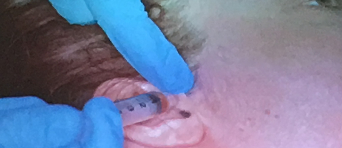
i-PRF | Pull in the syringe

The liquid is drawn into a syringe by penetrating the rubber top on the tube and by carefully suction fill the syringe.
i-PRF™ will stay as a liquid for approximately 12 minutes after
i-PRF | clotting and encapsulation
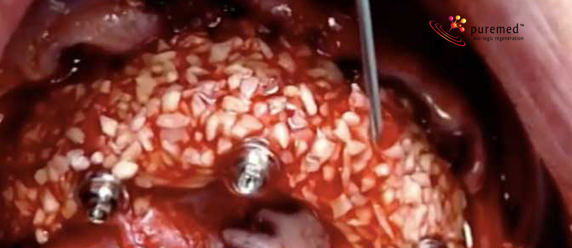
i-PRF™ injected onto a bone graft mix, where A-PRF™ membranes are mixed into the particles, will clot after few second and encapsulate the particles in a very nice way
i-PRF | 3 dimensional wound healing
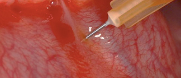
i-PRF™ allow wound healing specialists to add an injected “membrane” next to the A-PRF™ membranes placed on the wound bed.
i-PRF application | injections in knee joints

i-PRF™ injected into the knee joint show very promising results on pain reduction and in rebuild of cartilage.
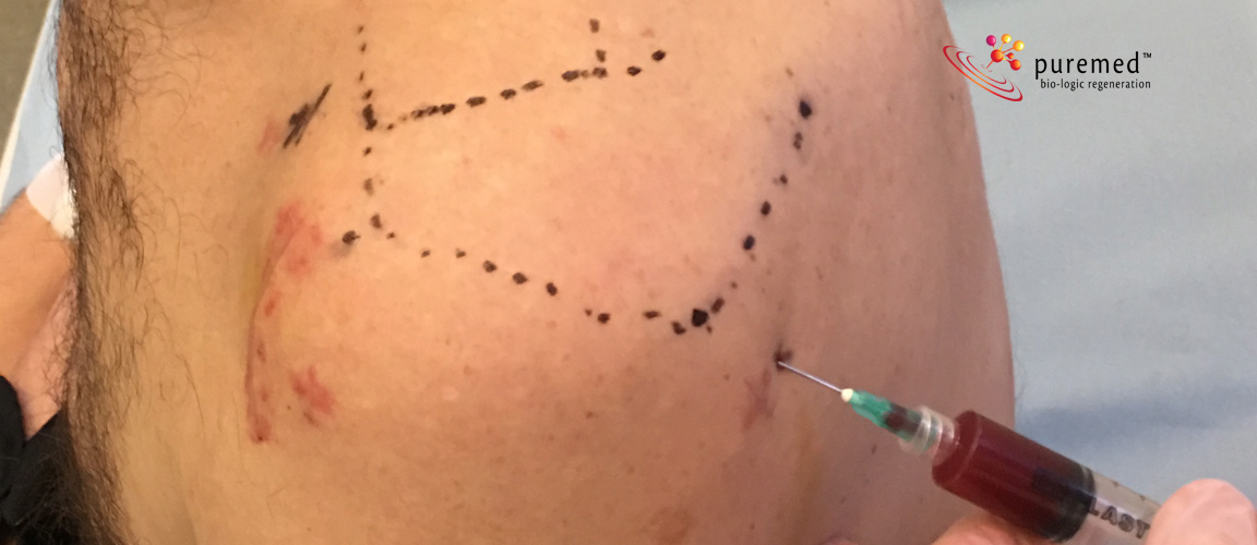
i-PRF application | injections in shoulders
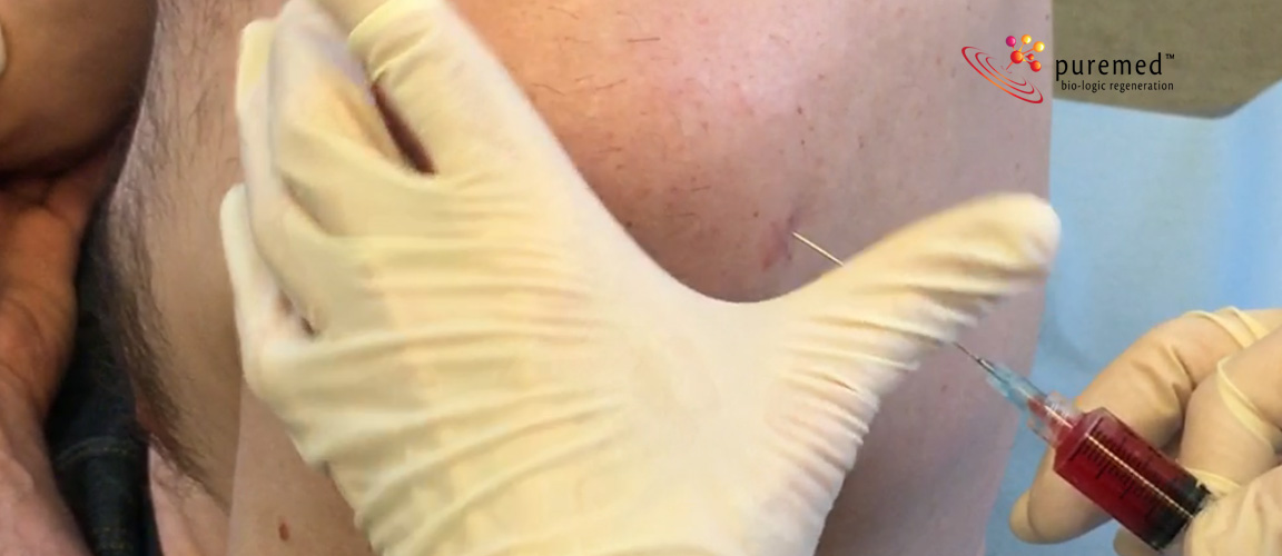
i-PRF™ injected into the the shoulder joint show very promising results on pain reduction.
i-PRF application | injections in jaw joint (TMJ)
i-PRF™ injected into the jaw joint (TMJ disorder) show very promising results on pain reduction.
-PRF™ | Team and litteratur

Dr. Joseph Choukroun
Dr. Joseph Choukroun
Received his diploma in 1979 in Montpellier, France. Specialist in anesthesiology in 1981, University of Montpellier. Fellowship of Pain Clinic, University of Strasbourg. Chief of staff of the Private Pain Clinic, Nice. President and creator of the SYFAC, international symposium on growth factors, Nice.
Inventor of the PRF technique. Author on several scientific and clinical papers for scientific journals. International Speaker.
International team
Dr. Joseph Choukroun is pushing the research on PRF together with scientists from the Laboratory of Clarion Research Group, Pennsylvania University (USA) and Repair-Lab, Institute of Pathology, Johannes Gutenberg University, Mainz (Germany) and Physicians around the world.
PRFs can be regarded as dense fibrin biomaterial with bio-mechanical properties. A high density fibrin clot can serve as a biological healing matrix by supporting cell migration and cytokine release, expanding the range of its potential applications greatly.
Source:Classification of platelet concentrates: from pure platelet-rich plasma (P-PRP) to leucocyte- and platelet-rich fibrin (L-PRF), David M. Dohan Ehrenfest, Lars Rasmusson and Tomas Albrektsson, Department of Biomaterials, Institute of Clinical Sciences, The Sahlgrenska Academy at University of Gothenburg, Sweden
A-PRF™ | Advantages of PRF over PRP
- No biochemical handling of blood.
- Simplified and cost-effective process.
- Use of bovine thrombin and anticoagulants not required.
- Favorable healing due to slow polymerization.
- More efficient cell migration and proliferation.
- PRF has supportive effect on immune system.
- PRF helps in hemostasis.
'PRF' 카테고리의 다른 글
| Advanced PRF, A-PRF의 우수성을 보여주는 논문 두 편! (0) | 2016.09.23 |
|---|---|
| PRF를 이용한 발치와 보존술, PRF socket preservation (0) | 2016.07.08 |
| 최신 PRF 논문 : A-PRF and A-PRF+ (0) | 2016.06.30 |
| PRF와 골아세포 (0) | 2016.06.29 |
| PRF와 치은퇴축 (0) | 2016.06.29 |